Life Extension Magazine®

How Vegetable Extracts Protect Against Cancer
Apigenin is a polyphenol found in vegetables such as parsley and celery. Data shows that apigenin can effectively starve cancer cells, guard DNA against toxins, and block malignant cell propagation.
Scientifically reviewed by: Dr. Amanda Martin, DC, in August 2023. Written by: Rita Haven.
Apigenin is a polyphenol found in vegetables such as parsley and celery. It is receiving increased attention as a low-cost nutrient to protect against common cancers.
What makes apigenin so fascinating is how it functions to starve cancer cells, promote cancer cell destruction, and protect cellular DNA against environmental toxins (that can result in future malignancies).
Compounds such as indole-3-carbinol (I3C) are found in cruciferous vegetables. These cruciferous compounds have been shown to work in complementary ways with apigenin (non-cruciferous) to combat cancer and other age-related diseases.
According to a study published in the International Journal of Oncology:
“Cancer prevention through diet may be largely achievable by increased consumption of fruits and vegetables. Considerable attention has been devoted to identifying plant-derived dietary agents which could be developed as promising chemopreventives. One such agent is apigenin.”1
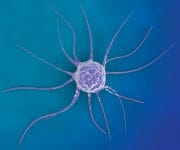
Apigenin Protects Cells from Cancer
Apigenin fights oxidative stress and inflammation—two factors that play a role in cancer development.2-9 Oxidative stress and inflammation generate DNA damage that can lead to uncontrolled proliferation of nonfunctioning cells, i.e., cancer.10,11 But that’s just one aspect of how apigenin functions.
Unique mechanisms of apigenin have led researchers to intensely evaluate it.12 What they’ve discovered is that apigenin has a host of other anticancer properties:
1. Apigenin Stops Cancer Cells from Replicating
Apigenin attacks cancer at a variety of stages and in different ways. At every step, apigenin seems to aggressively stop cancer’s various pathways. Apigenin has the ability to stop cancer cells from replicating, to reduce their invasiveness, and to slow their growth. Scientists believe this is largely related to its ability to shut down nuclear factor-kappa B (NF-kB).13,14 When activated, NF-kB leads to a flood of proinflammatory molecules that can promote tumor growth and the spread of cancer.15,16
In an animal study of nonmelanoma skin cancer, apigenin inhibited the production of inflammatory signaling molecules known to promote tumor proliferation.17
2. Apigenin Causes Cancer Cells to Die Off
In a cell study of chronic lymphocytic leukemia (a common malignancy in older adults), apigenin prevented the DNA mutations tied to cancer, while promoting the naturally-occurring cell death (apoptosis) that cancer cells otherwise evade.18
Apigenin may promote apoptosis by reactivating an important cancer-suppressor gene called p53.19 Inactivation of p53 is a common feature of cancer cells, which results in the cell’s loss of control over when to replicate and when to die naturally.20 By restoring p53 activity, apigenin essentially allows cancer cells to die a natural death.21
3. Apigenin Cuts off Cancer Cells’ Ability to Grow
In addition to causing cancer cells to die off naturally, studies have also shown that apigenin modulates a host of factors that can give cancer a promotional boost once it gets started.
Apigenin has repeatedly been shown to regulate the insulin-like growth factor signaling pathway known to promote the growth of prostate cancer cells when deregulated. Under the influence of apigenin, those cells quit their explosive growth and proceed to kill themselves off by apoptosis.12
4. Apigenin Starves Cancer Cells
Finally, apigenin appears capable of literally starving cancer cells into submission through several unrelated, but complementary, mechanisms.
First, apigenin suppresses the expression of a protein essential for transporting glucose into cancer cells.22,23
Apigenin further promotes this “energy crisis” by going after cancer cells’ mitochondria, the tiny intracellular power plants that generate energy. When mitochondria from human liver cancer cells were treated with apigenin, their membranes became leaky to the extent that it destroyed affected cancer cells.24
Based on the mechanisms by which apigenin thwarts cancer at every turn, new human studies are needed to further explore this impressive cancer-destroying polyphenol. The underlying mechanisms discussed here have been found to be effective in animal models of leukemia and the following solid tumor malignancies:
- Prostate14,25
- Larynx (voice box)22
- Leukemia18
- Liver (hepatocellular carcinoma)26
- Pancreatic9
- Skin17
- Breast27
What You Need to Know
 |
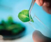
Health Benefits of Vegetable Extracts
- Vegetable extracts present in broccoli, celery, parsley, kale, Brussels sprouts, and many others—have long been revered for their health-promoting benefits.
- It is now recognized that four compounds, apigenin, I3C, DIM, and BITC, are responsible for the majority of that protection, and in very specific and complementary ways.
- All four have powerful cancer-protective effects, attacking and preventing malignancies through a multitude of overlapping mechanisms.
- Apigenin also has exceptional cardiovascular, metabolic, and neuroprotective properties, while I3C/DIM promote cardiovascular and liver health.
- Consumption of these plant compounds provides broad-spectrum protection against many of the most common symptoms of aging.
Apigenin Protects Brain Cells
Alzheimer’s and Parkinson’s are two common neurodegenerative diseases.28-30 Together, they cause untold misery for patients, their families, and their other caregivers. Numerous studies show apigenin’s ability to reduce some of the known contributors to neurodegeneration.
For example, apigenin fights excitotoxicity, the neuronal damage that occurs over a lifetime of intense brain cell stimulation.3,6 This is critical since excitotoxicity promotes brain cell death and dysfunction in both Alzheimer’s and Parkinson’s diseases.31,32
Apigenin also was shown to protect dopamine-producing cells of deep brain centers affected by Parkinson’s disease. This important action reduces neuroinflammation and the activation of inflammatory cells in the brain.33
When applied to brain cells in culture, apigenin protected those cells from toxicity induced by beta amyloid, the toxic “junk” protein found in abundance in the Alzheimer’s brain.34
Finally, given the importance of elevated blood sugar in the development of Alzheimer’s (it has been called “Type III diabetes” by some researchers), it is interesting to note that apigenin attenuates the cognitive decline seen in adult diabetic rats.4 Animals treated with apigenin have demonstrated improved learning and memory retention in a mouse model of Alzheimer’s disease.35
Apigenin, I3C, DIM, and BITC are compounds found in vegetables that offer wide-ranging protection against the factors that damage our DNA.
Apigenin Promotes Cardiometabolic Health

Oxidative stress and inflammation are deadly to heart and blood vessel cells. Inflammation/oxidation can also induce the kind of damage in liver and fat tissues that can promote weight gain, diabetes, and other metabolic changes that raise cardiovascular disease risk. Apigenin can contribute substantially to protecting against all of these effects.
At the most fundamental level, apigenin has been shown to prevent new cholesterol molecules from being synthesized in liver cells, which can reduce the amount of cholesterol in circulation.37 In a similar fashion, animal studies show that apigenin lowers blood sugar levels by decreasing insulin resistance, decreasing elevated insulin levels, and decreasing the formation of new glucose in the liver.37,38
Studies in diabetic rats reveal that apigenin can improve the function of the endothelial cells that line arteries and directly control blood flow and pressure.38
Preclinical studies show that apigenin helps protect the heart muscle against ischemia/reperfusion injury, the serious damage that occurs in the minutes to hours following a heart attack or stroke, when oxygen-starved (ischemic) tissue is suddenly flooded with oxygen-rich blood as circulation is restored (reperfusion).39-41
I3C: Complementary Tissue Protection from Cruciferous Vegetables
I3C (indole-3-carbinol) is a major component found in cruciferous vegetables. While I3C has many overlapping activities with apigenin, it also adds a substantial number of unique functions.42 Many of these benefits are also produced by 3,3’-diindolylmethane (DIM), a condensation product of I3C molecules.43,44
Through these unique functions, I3C and DIM provide complementary protection against cancer, heart disease, and more. Let’s take a look.
I3C/DIM: Unique Protection against Cancer
The most promising of I3C’s cancer-fighting properties has to do with its impact on enzymes that metabolize the sex hormones estrogen and testosterone.45
For example, I3C and DIM have been found to reduce a carcinogenic form of estrogen called 16-alpha-hydroxyestrone while boosting beneficial 2-hydroxyestrone. A large study of over 10,000 women showed that those with highest amounts of 2-hydroxyestrone compared to 16-alpha-hydroxyestrone had a remarkable 42% lower risk of breast cancer.46 Since hormone-dependent cancers such as breast and ovarian tumors thrive on unhealthy hormone balances, this is an important step in cancer prevention.42,47-49
There’s also considerable evidence that I3C modifies the function and expression of estrogen receptors on cells. Studies show that I3C downregulates the expression of estrogen receptor alpha, which is known to cause cancer-promoting cellular changes when it is overexpressed.49-52 Turning down estrogen receptor alpha allows greater influence for the protective estrogen receptor beta, which further reduces estrogen-dependent cancer risk.49
I3C/DIM Promote Cardiometabolic Health
I3C and DIM have multiple antiobesity effects.
Obese mice treated with I3C showed decreased body weight, fat accumulation, fat-mediated release of inflammatory cytokines, and fat infiltration by inflammatory cells—all of which produce obesity-associated health risks.53-56
I3C and DIM also reduce blood sugar, insulin, and markers of sugar-induced protein damage in mice on high-fat diets. These are effects that contribute to reduced atherosclerosis and cardiovascular disease risks.57,58
I3C/DIM Provide Wide-Ranging Liver Protection
I3C and DIM also have impressive liver-protective properties. These are partly due to their ability to suppress oxidative stress, in part through their activation of AMPK, and in part through their anti-inflammatory properties.59-60
These mechanisms of action protect the liver against fat accumulation, inflammatory changes, malignancies, and even fibrosis, the scarring and toughening that precedes liver failure.62
In fact, animal studies show that I3C and DIM can prevent fatty liver disease caused both by alcohol and a high-fat diet.59,60
BITC Offers Complementary Cancer Protection
Benzyl isothiocyanate (BITC) is another compound found in cruciferous vegetables that provides cancer protection that complements those of other cruciferous compounds.
One of its most startling properties is its ability to inhibit cancer stem cells. Many malignancies recur or fail to respond to treatment because of just a few cancer stem cells that can “hide out” and emerge after treatment is complete.
Both lab and animal studies show that BITC inhibits breast cancer stem cells, while also suppressing cell signaling molecules that contribute to the “stemness” of such cells.63-65 As a result, BITC has been found to be effective against numerous types of cancer.
For example, an animal study demonstrated that BITC reduced the number of prostate cancers in cancer-prone mice.67 BITC also prevents the stimulation of breast cancer growth induced by high-fat diets, prevents invasion and new blood vessel formation in brain and head and neck cancers, and induces cell death by apoptosis in lung cancers.67-70
Summary

Apigenin, I3C, DIM, and BITC are compounds found in vegetables that offer wide-ranging protection against the factors that damage our DNA.
These compounds work in a complementary way to protect our bodies against cancer, cardiovascular and metabolic diseases, neurodegeneration, and even liver disease.
Together, these botanical compounds represent the virtues of consuming more healthy vegetables in ones’ everyday diet.
For those who cannot consistently eat large amounts of celery, parsley and other vegetables, apigenin is available in low-cost multinutrient formulas that provide a wide variety of beneficial plant extracts.
If you have any questions on the scientific content of this article, please call a Life Extension® Wellness Specialist at 1-866-864-3027.
References
- Patel D, Shukla S, Gupta S. Apigenin and cancer chemoprevention: progress, potential and promise (review). Int J Oncol. 2007;30(1):233-45.
- Wang E, Chen F, Hu X, et al. Protective effects of apigenin against furan-induced toxicity in mice. Food Funct. 2014;5(8):1804-12.
- Han JY, Ahn SY, Kim CS, et al. Protection of apigenin against kainate-induced excitotoxicity by anti-oxidative effects. Biol Pharm Bull. 2012;35(9):1440-6.
- Mao XY, Yu J, Liu ZQ, et al. Apigenin attenuates diabetes-associated cognitive decline in rats via suppressing oxidative stress and nitric oxide synthase pathway. Int J Clin Exp Med. 2015;8(9): 15506-13.
- Nielsen SE, Young JF, Daneshvar B, et al. Effect of parsley (Petroselinum crispum) intake on urinary apigenin excretion, blood antioxidant enzymes and biomarkers for oxidative stress in human subjects. Br J Nutr. 1999;81(6):447-55.
- Balez R, Steiner N, Engel M, et al. Neuroprotective effects of apigenin against inflammation, neuronal excitability and apoptosis in an induced pluripotent stem cell model of Alzheimer’s disease. Sci Rep. 2016;6:31450.
- Smolinski AT, Pestka JJ. Modulation of lipopolysaccharide-induced proinflammatory cytokine production in vitro and in vivo by the herbal constituents apigenin (chamomile), ginsenoside Rb(1) (ginseng) and parthenolide (feverfew). Food Chem Toxicol. 2003;41(10):1381-90.
- Mascaraque C, Gonzalez R, Suarez MD, et al. Intestinal anti-inflammatory activity of apigenin K in two rat colitis models induced by trinitrobenzenesulfonic acid and dextran sulphate sodium. Br J Nutr. 2015;113(4):618-26.
- Wang YC, Huang KM. In vitro anti-inflammatory effect of apigenin in the Helicobacter pylori-infected gastric adenocarcinoma cells. Food Chem Toxicol. 2013;53:376-83.
- Klaunig JE, Kamendulis LM, Hocevar BA. Oxidative stress and oxidative damage in carcinogenesis. Toxicol Pathol. 2010;38(1):96-109.
- Kiraly O, Gong G, Olipitz W, et al. Inflammation-induced cell proliferation potentiates DNA damage-induced mutations in vivo. PLoS Genet. 2015;11(2):e1004901.
- Shukla S, Gupta S. Apigenin: a promising molecule for cancer prevention. Pharm Res. 2010;27(6):962-78.
- Cardenas H, Arango D, Nicholas C, et al. Dietary Apigenin Exerts Immune-Regulatory Activity in Vivo by Reducing NF-kappaB Activity, Halting Leukocyte Infiltration and Restoring Normal Metabolic Function. Int J Mol Sci. 2016;17(3):323.
- Shukla S, Kanwal R, Shankar E, et al. Apigenin blocks IKKalpha activation and suppresses prostate cancer progression. Oncotarget. 2015;6(31):31216-32.
- Tak PP, Firestein GS. NF-kappaB: a key role in inflammatory diseases. J Clin Invest. 2001;107(1):7-11.
- Karin M. NF-kappaB as a critical link between inflammation and cancer. Cold Spring Harb Perspect Biol. 2009;1(5):a000141.
- Kiraly AJ, Soliman E, Jenkins A, et al. Apigenin inhibits COX-2, PGE2, and EP1 and also initiates terminal differentiation in the epidermis of tumor bearing mice. Prostaglandins Leukot Essent Fatty Acids. 2016;104:44-53.
- Hashemi M, Nouri Long M, Entezari M, et al. Anti-mutagenic and pro-apoptotic effects of apigenin on human chronic lymphocytic leukemia cells. Acta Med Iran. 2010;48(5):283-8.
- King JC, Lu QY, Li G, et al. Evidence for activation of mutated p53 by apigenin in human pancreatic cancer. Biochim Biophys Acta. 2012;1823(2):593-604.
- Lane DP, Cheok CF, Lain S. p53-based cancer therapy. Cold Spring Harb Perspect Biol. 2010;2(9):a001222.
- Zheng PW, Chiang LC, Lin CC. Apigenin induced apoptosis through p53-dependent pathway in human cervical carcinoma cells. Life Sci. 2005;76(12):1367-79.
- Bao YY, Zhou SH, Lu ZJ, et al. Inhibiting GLUT-1 expression and PI3K/Akt signaling using apigenin improves the radiosensitivity of laryngeal carcinoma in vivo. Oncol Rep. 2015;34(4):1805-14.
- Lee YM, Lee G, Oh TI, et al. Inhibition of glutamine utilization sensitizes lung cancer cells to apigenin-induced apoptosis resulting from metabolic and oxidative stress. Int J Oncol. 2016;48(1):399-408.
- Seydi E, Rasekh HR, Salimi A, et al. Selective Toxicity of Apigenin on Cancerous Hepatocytes by Directly Targeting their Mitochondria. Anticancer Agents Med Chem. 2016.
- Babcook MA, Gupta S. Apigenin Modulates Insulin-like Growth Factor Axis: Implications for Prevention and Therapy of Prostate Cancer. Curr Drug Targets. 2012.
- Hu XY, Liang JY, Guo XJ, et al. 5-Fluorouracil combined with apigenin enhances anticancer activity through mitochondrial membrane potential (DeltaPsim)-mediated apoptosis in hepatocellular carcinoma. Clin Exp Pharmacol Physiol. 2015;42(2):146-53.
- Mafuvadze B, Cook M, Xu Z, et al. Effects of dietary apigenin on tumor latency, incidence and multiplicity in a medroxyprogesterone acetate-accelerated 7,12-dimethylbenz(a)anthracene-induced breast cancer model. Nutr Cancer. 2013;65(8):1184-91.
- Checkoway H, Lundin JI, Kelada SN. Neurodegenerative diseases. IARC Sci Publ. 2011(163):407-19.
- Available at: https://www.aan.com/uploadedFiles/Website_Library_Assets/Documents/6.Public_Policy/1.Stay_Informed/4.Public_Policy_Resources/compell.pdf. Accessed October 13, 2016.
- Available at: https://www.niehs.nih.gov/research/supported/health/neurodegenerative/index.cfm. Accessed October 13, 2016.
- Koutsilieri E, Riederer P. Excitotoxicity and new antiglutamatergic strategies in Parkinson’s disease and Alzheimer’s disease. Parkinsonism Relat Disord. 2007;13 Suppl 3:S329-31.
- Hugon J, Vallat JM, Dumas M. Role of glutamate and excitotoxicity in neurologic diseases. Rev Neurol (Paris). 1996;152(4):239-48.
- Liu W, Kong S, Xie Q, et al. Protective effects of apigenin against 1-methyl-4-phenylpyridinium ioninduced neurotoxicity in PC12 cells. Int J Mol Med. 2015;35(3):739-46.
- Zhao L, Wang JL, Wang YR, et al. Apigenin attenuates copper-mediated beta-amyloid neurotoxicity through antioxidation, mitochondrion protection and MAPK signal inactivation in an AD cell model. Brain Res. 2013;1492:33-45.
- Zhao L, Wang JL, Liu R, et al. Neuroprotective, anti-amyloidogenic and neurotrophic effects of apigenin in an Alzheimer’s disease mouse model. Molecules. 2013;18(8):9949-65.
- Wong TY, Lin SM, Leung LK. The flavone apigenin blocks nuclear translocation of sterol regulatory element-binding protein-2 in the hepatic cells WRL-68. Br J Nutr. 2015;113(12):1844-52.
- Jung UJ, Cho YY, Choi MS. Apigenin Ameliorates Dyslipidemia, Hepatic Steatosis and Insulin Resistance by Modulating Metabolic and Transcriptional Profiles in the Liver of High-Fat Diet-Induced Obese Mice. Nutrients. 2016;8(5).
- Ren B, Qin W, Wu F, et al. Apigenin and naringenin regulate glucose and lipid metabolism, and ameliorate vascular dysfunction in type 2 diabetic rats. Eur J Pharmacol. 2016;773:13-23.
- Yang X, Yang J, Hu J, et al. Apigenin attenuates myocardial ischemia/reperfusion injury via the inactivation of p38 mitogenactivated protein kinase. Mol Med Rep. 2015;12(5):6873-8.
- Ferrari R, Pepi P, Ferrari F, et al. Metabolic derangement in ischemic heart disease and its therapeutic control. Am J Cardiol. 1998;82(5a):2k-13k.
- Hu J, Li Z, Xu LT, et al. Protective effect of apigenin on ischemia/reperfusion injury of the isolated rat heart. Cardiovasc Toxicol. 2015;15(3):241-9.
- Aggarwal BB, Ichikawa H. Molecular targets and anticancer potential of indole-3-carbinol and its derivatives. Cell Cycle. 2005;4(9):1201-15.
- Xue L, Pestka JJ, Li M, et al. 3,3’-Diindolylmethane stimulates murine immune function in vitro and in vivo. J Nutr Biochem. 2008;19(5):336-44.
- Rahman KM, Ali S, Aboukameel A, et al. Inactivation of NF-kappaB by 3,3’-diindolylmethane contributes to increased apoptosis induced by chemotherapeutic agent in breast cancer cells. Mol Cancer Ther. 2007;6(10):2757-65.
- Wang TT, Schoene NW, Milner JA, et al. Broccoli-derived phytochemicals indole-3-carbinol and 3,3’-diindolylmethane exerts concentration-dependent pleiotropic effects on prostate cancer cells: comparison with other cancer preventive phytochemicals. Mol Carcinog. 2012;51(3):244-56.
- Muti P1, Bradlow HL, Micheli A, et al. Estrogen metabolism and risk of breast cancer: a prospective study of the 2:16alpha-hydroxyestrone ratio in premenopausal and postmenopausal women. Epidemiology. 2000 Nov;11(6):635-40.
- Higdon JV, Delage B, Williams DE, et al. Cruciferous vegetables and human cancer risk: epidemiologic evidence and mechanistic basis. Pharmacol Res. 2007;55(3):224-36.
- Rogan EG. The natural chemopreventive compound indole-3-carbinol: state of the science. In Vivo. 2006;20(2):221-8.
- Weng JR, Tsai CH, Kulp SK, et al. Indole-3-carbinol as a chemopreventive and anti-cancer agent. Cancer Lett. 2008;262(2):153-63.
- Kim YS, Milner JA. Targets for indole-3-carbinol in cancer prevention. J Nutr Biochem. 2005;16(2):65-73.
- Marconett CN, Sundar SN, Tseng M, et al. Indole-3-carbinol downregulation of telomerase gene expression requires the inhibition of estrogen receptor-alpha and Sp1 transcription factor interactions within the hTERT promoter and mediates the G1 cell cycle arrest of human breast cancer cells. Carcinogenesis. 2011;32(9):1315-23.
- Marconett CN, Singhal AK, Sundar SN, et al. Indole-3-carbinol disrupts estrogen receptor-alpha dependent expression of insulin-like growth factor-1 receptor and insulin receptor substrate-1 and proliferation of human breast cancer cells. Mol Cell Endocrinol. 2012;363(1-2):74-84.
- Chang HP, Wang ML, Chan MH, et al. Antiobesity activities of indole-3-carbinol in high-fat-diet-induced obese mice. Nutrition. 2011;27(4):463-70.
- Chang HP, Wang ML, Hsu CY, et al. Suppression of inflammation-associated factors by indole-3-carbinol in mice fed high-fat diets and in isolated, co-cultured macrophages and adipocytes. Int J Obes (Lond). 2011;35(12):1530-8.
- Choi Y, Kim Y, Park S, et al. Indole-3-carbinol prevents diet-induced obesity through modulation of multiple genes related to adipogenesis, thermogenesis or inflammation in the visceral adipose tissue of mice. J Nutr Biochem. 2012;23(12):1732-9.
- Wang ML, Lin SH, Hou YY, et al. Suppression of Lipid Accumulation by Indole-3-Carbinol Is Associated with Increased Expression of the Aryl Hydrocarbon Receptor and CYP1B1 Proteins in Adipocytes and with Decreased Adipocyte-Stimulated Endothelial Tube Formation. Int J Mol Sci. 2016;17(8).
- Jayakumar P, Pugalendi KV, Sankaran M. Attenuation of hyperglycemia-mediated oxidative stress by indole-3-carbinol and its metabolite 3, 3’- diindolylmethane in C57BL/6J mice. J Physiol Biochem. 2014;70(2):525-34.
- Poornima J, Mirunalini S. Regulation of carbohydrate metabolism by indole-3-carbinol and its metabolite 3,3’-diindolylmethane in high-fat diet-induced C57BL/6J mice. Mol Cell Biochem. 2014;385 (1-2):7-15.
- Guo Y, Wu XQ, Zhang C, et al. Effect of indole-3-carbinol on ethanol-induced liver injury and acetaldehyde-stimulated hepatic stellate cells activation using precision-cut rat liver slices. Clin Exp Pharmacol Physiol. 2010;37(12):1107-13.
- Choi Y, Yanagawa Y, Kim S, et al. Involvement of SIRT1-AMPK signaling in the protective action of indole-3-carbinol against hepatic steatosis in mice fed a high-fat diet. J Nutr Biochem. 2013;24(7):1393-400.
- Busbee PB, Nagarkatti M, Nagarkatti PS. Natural indoles, indole-3-carbinol (I3C) and 3,3’-diindolylmethane (DIM), attenuate staphylococcal enterotoxin B-mediated liver injury by downregulating miR-31 expression and promoting caspase-2-mediated apoptosis. PLoS One. 2015;10(2):e0118506.
- Wang SQ, Cheng LS, Liu Y, et al. Indole-3-Carbinol (I3C) and its Major Derivatives: Their Pharmacokinetics and Important Roles in Hepatic Protection. Curr Drug Metab. 2016;17(4):401-9.
- Kim SH, Sehrawat A, Singh SV. Dietary chemopreventative benzyl isothiocyanate inhibits breast cancer stem cells in vitro and in vivo. Cancer Prev Res (Phila). 2013;6(8):782-90.
- Rao CV. Benzyl isothiocyanate: double trouble for breast cancer cells. Cancer Prev Res (Phila). 2013;6(8):760-3.
- Kim SH, Singh SV. The role of polycomb group protein Bmi-1 and Notch4 in breast cancer stem cell inhibition by benzyl isothiocyanate. Breast Cancer Res Treat. 2015;149(3):681-92.
- Cho HJ, Lim do Y, Kwon GT, et al. Benzyl Isothiocyanate Inhibits Prostate Cancer Development in the Transgenic Adenocarcinoma Mouse Prostate (TRAMP) Model, Which Is Associated with the Induction of Cell Cycle G1 Arrest. Int J Mol Sci. 2016;17(2):264.
- Liu BN, Yan HQ, Wu X, et al. Apoptosis induced by benzyl isothiocyanate in gefitinib-resistant lung cancer cells is associated with Akt/MAPK pathways and generation of reactive oxygen species. Cell Biochem Biophys. 2013;66(1):81-92.
- Zhu Y, Zhang L, Zhang GD, et al. Potential mechanisms of benzyl isothiocyanate suppression of invasion and angiogenesis by the U87MG human glioma cell line. Asian Pac J Cancer Prev. 2014;15(19):8225-8.
- Kim M, Cho HJ, Kwon GT, et al. Benzyl isothiocyanate suppresses high-fat diet-stimulated mammary tumor progression via the alteration of tumor microenvironments in obesity-resistant BALB/c mice. Mol Carcinog. 2015;54(1):72-82.
- Wolf MA, Claudio PP. Benzyl isothiocyanate inhibits HNSCC cell migration and invasion, and sensitizes HNSCC cells to cisplatin. Nutr Cancer. 2014;66(2):285-94.
Wellness
Specialists
1-800-226-2370 - This service is FREE
7:30 AM - 12 AM (ET) Mon-Fri | 9 AM - 12 AM (ET) Sat-Sun